CR-39 Plastic Nuclear Track Detector
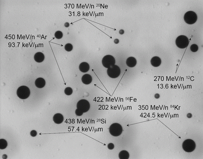
Columbia Resin No. 39, or CR-39, Plastic Nuclear Track Detector (PNTD) is sensitive to protons of energy ≤ 14 MeV, alpha particles of energy ≤ 100 MeV, and heavy ions of all energies. Neutrons of energy between 500 keV and 20 MeV can be detected via an enlarged latent damage trail, or "track," from recoil protons formed as a result of interactions between neutrons and the hydrogen nuclei of the detector. CR-39 PNTD measures Linear Energy Transfer (LET) fluence, dose, and dose equivalent spectrum from particles of LET∞H2O ≥ 5 keV/µm, examples of which are shown in Figure 1.
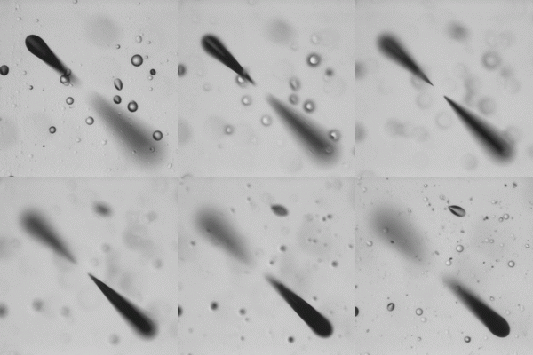
It can also measure the charge spectrum of secondary fragments when used in laboratory testing during exposure to mono-energetic heavy ion beams. CR-39 PNTD consists of thin layers of thermoset polymer. The entire dosimeter is often smaller than a credit card. When a charged particle passes through the detector, it breaks the chemical bonds of the polymer, leaving a “latent damage trail” along the particle’s trajectory. An example of an enlarged latent damage trail, which is informally referred to as a "track," is shown in Figure 2.
In practice, CR-39 PNTD is very similar to photographic film in the way it is used. After the detector has been exposed to a radiation field, either naturally present or created in the laboratory, it must be "developed" in order to make the detector readable by optical means. CR-39 is developed by chemically etching each detector in a highly alkaline solution that preferentially attacks the latent damage trail, leaving a conical pit at the intersection of the particle’s trajectory and the detector surface. The result of this chemical processing is shown in Figure 3.
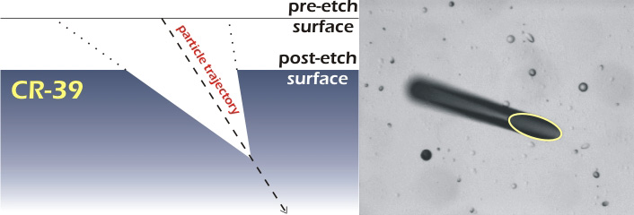 Figure 3: By chemically removing material from the detector, the latent damage trails are
enlarged, thereby becoming what are called "tracks." In this way optical read-out
of the CR-39 is possible.
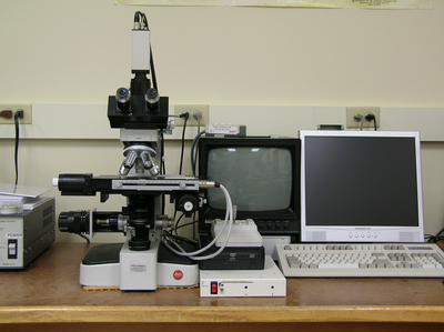
The dimensions of the elliptical opening of the conical track are proportional to the LET of the charged particle that created the latent damage trail. The etched detector is then read-out using a dedicated automatic microscope system, shown in Figure 4.
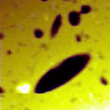
The limit of optical microscopy is approximately 6 μm. To accurately resolve features smaller than this size we must use an atomic force microscope (AFM). The AFM uses a very small, very sharp tip to physically trace the features of a surface. The radius of curvature for a new AFM tip is typically 20 nm. The resulting topographic data is then displayed using a false color map to show the variations in surface height. We can then use various methods to measure the physical dimensions of the etched nuclear tracks. This allows us to calculate the properties of the particle that caused the track. CR-39, in conjunction with the AFM, is one of only a few methods by which short-range high-LET recoils produced in nuclear target fragmentation interactions can be directly measured. Figure 5 shows an example of a 10 µm × 10 µm AFM scan of CR-39 PNTD registering the track of a heavy nuclear recoil.
In Figure 5, dark areas are deeper than the yellow areas. The detector scanned in this image was placed behind a gold target and exposed to a 1 GeV proton beam. The CR-39 material records the passage of protons, knock-out particles, and heavy nuclear recoil fragments. This research has applications in space dosimetry and proton cancer therapy.

